We provide genetic testing services for the following Rare diseases:

Facioscapulohumeral Muscular Dystrophy (FSHD)

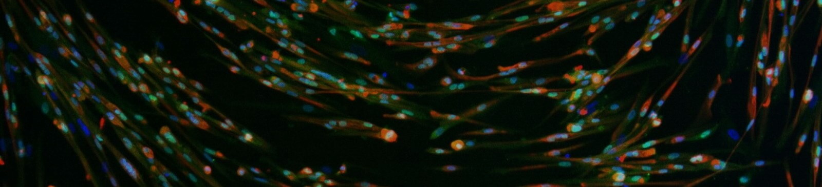
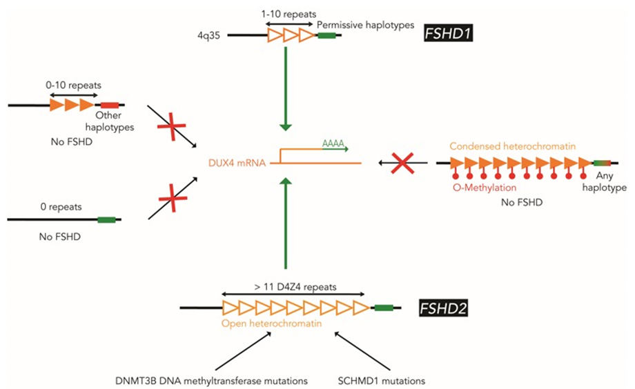
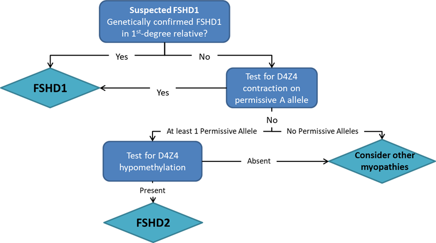
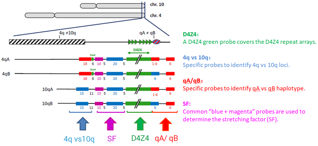
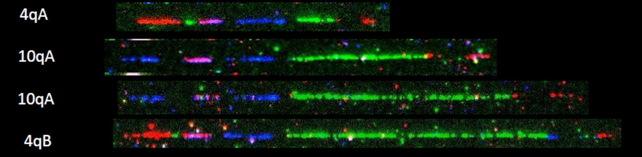
Pangenia Genomics provides the genetic testing of FHSD1 based on the Molecular Combing and Genomic Morse Code technology. This method uses specific fluorescent probes to hybridize with genomic DNA molecules (extracted from fresh blood samples) linearly stretched on coverslips. The fluorescent signals can then be visualized to distinguish the length (number of D4Z4 repeat units) and the type (haplotype A or B) of the D4Z4 loci. Most affected individuals have fewer than 10 repeats and the 4qA allele.

Learn More

Duchenne Muscular Dystrophy (DMD) and Becker Muscular Dystrophy (BMD)

Pangenia Genomics offers deletion/duplication analysis to accurately detect the number of copies of the DMD gene, i.e. find out if the gene or part of the gene is deleted or duplicated. It can be used for carrier screening as well.

Learn More

Spinal Muscular Atrophy (SMA)

Pangenia Genomics offers deletion/duplication analysis in order to accurately detect deletions and duplications in the SMN1 gene. It can be used for carrier screening as well.

Learn More

Charcot-Marie-Tooth (CMT) disease

Pangenia Genomics offers an in vitro diagnostic (IVD) test for the detection of deletions or duplications in the PMP22, MPZ and GJB1 genes for the diagnosis of Charcot-Marie-Tooth disease type 1 (CMT1) disease. The test can be used to confirm the cause and diagnosis for hereditary neuropathy with liability to pressure palsies (HNPP) as well.

Learn More

Other Rare Neuromuscular Disorders

Learn More

What is FSHD?

Facioscapulohumeral muscular dystrophy, or FSHD, is a genetic disorder affecting the skeletal muscles. It typically starts in the early adolescence with the weakness of muscles in the face (facio), shoulders (scapula), upper arms (humerus), and legs. Weakness is slowly progressive and can spread to any muscle. Severity is highly variable among individuals. Approximately 20% of affected individuals eventually require a wheelchair. Life expectancy is not shortened. FSHD is estimated to affect between 4 and 10 individuals per 100,000 people.

Symptoms of FSHD include (but not always):

  • Unable to whistle or pucker lips
  • Unable to sip through a straw
  • Eyes dont close fully during sleep
  • Shoulder blades that wing out
  • Hard to raise the arms above shoulder
  • Difficulty with sit-ups and pull-ups
  • Weakness in hands and fingers
  • Trouble with rising from a chair
  • Difficulty in walking; a waddling gait
  • Foot droop
  • Protuberant abdomen
  • Curved spine
  • Chronic fatigue
  • Muscle pain

Genetic Cause and Inheritance

FSHD is caused by the abnormal expression of the DUX4 gene, located in the D4Z4 region near the end of chromosome 4 at the 4q35 location. Normally, the DNA in the D4Z4 region includes 11-100 repeated segments of DNA and is hypermethylated, repressing the expression of the DUX4 gene. In FSHD, the expression of DUX4 is reactivated, with two underlying genetic mechanisms.

FSHD1

FSHD1 accounts for 95% of FSHD cases. In individuals with FSHD1, the D4Z4 array is contracted to 1 to 10 units, leading to the relaxation and hypomethylation of chromatin, and the toxic expression of DUX4 gene in cells and tissues where it is usually not produced.
The contraction of the D4Z4 array by itself is not sufficient to induce DUX4 expression. Immediately distal to the D4Z4 region, there is a polyadenylation site required for the stable expression of the DUX4 gene. The sequence has two variations, called type 4qA and 4qB. Only the 4qA variant in combination with the contracted D4Z4 repeat array can result in FSHD. DUX4 cannot be stably expressed with 4qB regardless of the length of D4Z4 repeat array. In addition, an almost identical D4Z4 array is also present on chromosome 10q26, the contraction of which does not result in the expression of DUX4 gene due to mutations in its polyadenylation site, and thus does not cause FSHD.
FSHD1 is inherited in an autosomal dominant manner. Offspring of an affected parent have a 50% chance of inheriting FSHD. About 70%-90% of FSHD1-affected individuals inherited the disease-causing D4Z4 array contraction from a parent, and about 10%-30% of affected individuals get FSHD due to de novo mutation.

FSHD2

FSHD2 is clinically identical to FSHD1 but genetically different. In FSHD2, the length of the D4Z4 repeat array on chromosome 4q35 is in normal range. Around 80% of FSHD2 individuals carry a heterozygous mutation in the SMCHD1 gene, and some FSHD2 individuals have mutations in the DNMT3B gene. These mutations are associated with the hypomethylation and relaxation of chromatin in the D4Z4 repeat arrays on both chromosome 4q35 and 10q26. In combination with the 4qA allele, DUX4 is expressed from the D4Z4 repeat array on chromosome 4q35, leading to FSHD. FSHD2 is inherited in a digenic manner.

Figure 1: Schematic illustration of FSHD molecular pathogenesis

Diagnosis

FSHD may be diagnosed through a thorough clinical examination, identification of characteristic physical findings, a complete individual and family history, and genetic testing (Figure 2). It usually starts with the testing of FHSD1, which accounts for 95% of the FSHD cases. For many years, the genetic testing of FSHD1 used Southern blot, which is labour and time-intensive, and just provides an estimate of the number of D4Z4 repeats. Pangenia Genomics is the first in Hong Kong to provide the genetic test of FHSD1 based on the Molecular Combing and Genomic Morse Code technology developed by Genomic Vision (http://www.genomicvision.com/products/genetic-tests/fshd/). This method uses specific fluorescent probes to hybridize with genomic DNA molecules (extracted from fresh blood samples) linearly stretched on coverslips (Figure 3). The fluorescent signals can then be visualized to distinguish the length (number of D4Z4 repeat units) and the type (haplotype A or B) of the D4Z4 loci (Figure 4). Most affected individuals have fewer than 10 repeats and the 4qA allele.

Genetic testing for mutations in the SMCHD1 and DNMT3B gene may be indicated if the D4Z4 region on chromosome 4 is not contracted.

Figure 2: Recommended diagnostic flowchart for FSHD

Figure 3: FSHD Genomic Morse Code (GMC) Design

Figure 4: Examples of hybridization signals

Treatment

Currently there is no treatment for FSHD, but several therapies are under investigation. Genetic treatments such as RNAi treatment to silence DUX4 have been evaluated in preclinical studies, though no human trials are currently underway. Losmapimod has been shown in preclinical studies to reduce DUX4 expression, and Phase II trials are currently enrolling for FSHD. Another FSHD treatment candidate GBC0905 is in preclinical studies (https://myocea.com/fshd).

References

[1] What is FSHD? FSHD Society.
https://www.fshdsociety.org/what-is-fshd/
[2] GeneReviews® Facioscapulohumeral Muscular Dystrophy
https://www.ncbi.nlm.nih.gov/books/NBK1443/
[3] NORD-Rare Disease Database: Facioscapulohumeral Muscular Dystrophy
https://rarediseases.org/rare-diseases/facioscapulohumeral-muscular-dystrophy/

What is DMD & BMD?

Dystrophinopathies are diseases that are caused due to mutation in the dystrophin-coding gene, including Duchenne Muscular Dystrophy (DMD) and Becker Muscular Dystrophy (BMD). They are one of the most common neuromuscular diseases in Hong Kong. According to a local study, the prevalence of dystrophinopathies (including DMD and BMD) in Hong Kong is around 1.03 per 10 000 males. The global prevalence is 7.1 per 100,000 males.

DMD is one of the most severe types of muscular dystrophies. It is an early onset disease which causes progressive muscle degeneration. Patients start to have symptoms since childhood.

Symptoms of DMD

  • Falling frequently
  • Difficulty climbing stairs, walking and running
  • Difficulty performing daily activities
  • Muscle pain
  • In more severe cases, patients may have cardiac and respiratory problems as well, which may lead to death.
Quality of life of patients is affected and lifespan is shortened. Even though the life expectancy has increased with the help of treatment and management, most affected only survive until young adulthood.
BMD is a milder form of DMD, and is caused by the same gene but different mutations. Symptoms begin at a later age, and progressive muscle degeneration is slower. BMD patients generally have stronger muscles than DMD patients, and may survive till their 40s.

Inheritance

DMD and BMD are inherited diseases that can be passed on to the next generation through carriers of the defective gene. As they are X-linked recessive diseases, two copies of the defective gene (located on the X-chromosome) are required for the disease to develop. Therefore, mainly males are affected. Female cases of DMD patients are rare but they can be carriers of the defective gene and have a 50% chance of passing on the defective gene to their children.

Mutations and Testing

DMD is caused by mutations in the DMD gene, which encodes the dystrophin protein. The most common mutations include large deletions and large duplications. Other mutations include point mutations and insertions.

DMD can be tested by numerous methods, including blood tests, muscle biopsy, CGH-array, multiplex PCR, direct sequencing, and deletion/duplication analysis. Pangenia Genomics offers deletion/duplication analysis accurately detect the number of copies of the DMD gene, i.e. find out if the gene or part of the gene is deleted or duplicated. It can be used for carrier screening as well.

Management and Treatment

  • Physiotherapy
  • Nutrition management
  • Surgery (for cardiac problems)
  • Drugs to delay disease progression - Glucocorticoids (Prednisone / Deflazacort)
  • FDA approved drug Eteplirsen for exon 51 skipping, so that the body is able to produce shorter but functional dystrophin hereby reducing the severity of the disease.
  • Drug (Ataluren – licensed in Europe) to treat DMD cases with nonsense mutation.

References

[1] Crisafulli S, Sultana J, Fontana A, Salvo F, Messina S, Trifirò G. Global epidemiology of Duchenne muscular dystrophy: an updated systematic review and meta-analysis. Orphanet Journal of Rare Diseases. 2020;15(1).
[2] Chan S, Lo I, Cherk S, Cheng W, Fung E, Yeung W et al. Prevalence and Characteristics of Chinese Patients With Duchenne and Becker Muscular Dystrophy. Child Neurology Open. 2015;2(2):2329048X1558534.
[3] Landrum Peay H, Fischer R, Tzeng J, Hesterlee S, Morris C, Strong Martin A et al. Gene therapy as a potential therapeutic option for Duchenne muscular dystrophy: A qualitative preference study of patients and parents. PLOS ONE. 2019;14(5):e0213649.
[4] Duchenne Muscular Dystrophy - NORD (National Organization for Rare Disorders) [Internet]. NORD (National Organization for Rare Disorders). 2020 [cited 12 November 2020]. Available from:
https://rarediseases.org/rare-diseases/duchenne-muscular-dystrophy/#:~:text=Duchenne%20muscular%20dystrophy%20(DMD)%20is,and%20six%20years%20of%20age
[5] Duchenne muscular dystrophy | Genetic and Rare Diseases Information Center (GARD) – an NCATS Program [Internet]. Rarediseases.info.nih.gov. 2020 [cited 12 November 2020]. Available from:
https://rarediseases.info.nih.gov/diseases/6291/duchenne-muscular-dystrophy
[6] Queen Mary Hospital and The Duchess of Kent Children's Hospital at Sandy Bay, Hong Kong Paediatric Neuromuscular Diagnostic and Management Program [Internet]. Paed.hku.hk. 2020 [cited 12 November 2020]. Available from:
http://paed.hku.hk/website/nmd/DMD.html

What is SMA?

Spinal Muscular Atrophy (SMA) is a neuromuscular disorder. It affects the survival motor neuron 1 gene (SMN1) which encodes the SMN protein. Around 95% of affected patients have deletion of exon 7 in both copies of the gene. Other mutations include point mutations. Prevalence of SMA is around 1 in 6,000 – 10,000 live births.

There is an SMN2 gene which is highly similar to SMN1, differing by a nucleotide in exon 7. Due to the difference, SMN2 has lower efficiency than SMN1, hence can produce small amounts of SMN protein but cannot fully replace SMN1. The SMN2 gene can reflect the severity of the disease in SMA patients since different people have different numbers of SMN2 copies.

Main Symptoms

  • Muscular atrophy
  • Hypotonia
  • Muscle weakness

Inheritance

SMA is an autosomal recessive disorder. If an individual has two defective genes, they will be affected by SMA. If an individual only has 1 defective gene, they are carriers. If a parent is a carrier, they have 50% chance of passing the defective gene to their children. If both parents are carriers, they have 25% chance of having a child affected with SMA.

Types of SMA and their symptoms

SMA is divided into types based on the age of onset and the severity of symptoms:

Type 0

  • rarest type of SMA
  • prenatal onset
  • the affected usually cannot survive beyond 6 months after birth

Type 1

  • the most severe after type 0
  • early onset (before 6 months old)
  • cannot sit
  • difficulty swallowing
  • usually survive till 2 years old (respiratory failure)

Type 2

  • the most severe after type 0
  • early onset (before 6 months old)
  • cannot sit
  • difficulty swallowing
  • usually survive till 2 years old (respiratory failure)

Type 3

  • onset: 18 months to 18 year
  • able to walk
  • muscle pain
  • joint pain
  • survive till adulthood

Type 4

  • around 1% cases
  • least severe
  • adult-onset
  • able to walk
  • may not have symptoms
  • normal lifespan

Diagnosis

It is important to get a diagnosis as early as possible in order to start proper management of the condition. Tests include muscle biopsy, EMG tests and genetic testing. Pangenia Genomics offers deletion/duplication analysis in order to accurately detect deletions and duplications in the SMN1 gene. It can be used for carrier screening as well.

Management and Treatment

Treatment can help slow progression of disease and may increase lifespan.

  • Nutrition management
  • Physical therapy
  • FDA approved treatments:
    • nusinersen – targets SMN2
    • onasemnogene abeparvovec-xioi - SMN1 gene replacement
    • risdiplam – targets SMN2 (newly approved)

References

[1] Queen Mary Hospital and The Duchess of Kent Children's Hospital at Sandy Bay, Hong KongPaediatric Neuromuscular Diagnostic and Management Program [Internet]. Paed.hku.hk. 2020 [cited 12 November 2020]. Available from:
http://paed.hku.hk/website/nmd/SMA.html
[2] Spinal Muscular Atrophy - NORD (National Organization for Rare Disorders) [Internet]. NORD (National Organization for Rare Disorders). 2020 [cited 12 November 2020]. Available from:
https://rarediseases.org/rare-diseases/spinal-muscular-atrophy/
[3] Spinal muscular atrophy | Genetic and Rare Diseases Information Center (GARD) – an NCATS Program [Internet]. Rarediseases.info.nih.gov. 2020 [cited 12 November 2020]. Available from:
https://rarediseases.info.nih.gov/diseases/7674/spinal-muscular-atrophy/
[4] Kolb S, Kissel J. Spinal Muscular Atrophy. Neurologic Clinics. 2015;33(4).
[5] Prior T, Swoboda K, Scott H, Hejmanowski A. HomozygousSMN1 deletions in unaffected family members and modification of the phenotype bySMN2. American Journal of Medical Genetics. 2004;130A(3):307-310.

Charcot-Marie-Tooth (CMT) disease

Charcot-Marie-Tooth (CMT) disease is a group of disorders in which the motor and/or sensory peripheral nerves are affected, resulting in muscle weakness and atrophy, as well as sensory loss. CMT is the most common form of inherited peripheral neuropathies. The nerve cells in individuals with this disorder are not able to send electrical signals properly because of abnormalities in the nerve axon or abnormalities in the insulation (myelin) around the axon. Specific gene mutations are responsible for the abnormal function of the peripheral nerves. Prevalence of CMT and related disorders has been estimated to be between 1:2500 and 1:1214.

Main Symptoms

Clinical symptoms of CMT include distal muscle weakness and atrophy, sensory loss, depressed tendon reflexes and high-arched feet (pes cavus). These manifestations occur first in the distal legs and later in the hands. At present, more than 80 genes are known to be associated with different types of CMT.

Inheritance

More than 80 genes are known to be associated with different types of CMT. CMT diseases can be inherited in an autosomal dominant, autosomal recessive or X-linked manner.

Diagnosis

CMT type 1 (CMT1) is a slowly demyelinating peripheral neuropathy and several subtypes exist. CMT1A accounts for approximately 70-80% of all CMT1 cases, which is mainly caused by an approximately 1.5 Mb duplication on chromosome 17p, including the peripheral myelin protein 22 (PMP22) gene and the flanking regions. In addition to duplication, CMT1A can be caused by point mutations in the gene PMP22 as well.
Increased PMP22 gene dosage leads to altered nerve conduction velocity, which is the main cause of the clinical manifestations in CMT1A.

Another subtype of CMT disease is hereditary neuropathy with liability to pressure palsies (HNPP) and PMP22 is the only gene known to be associated with HNPP which is characterized by repeated focal pressure neuropathies such as carpal tunnel syndrome and peroneal palsy with foot drop. A deletion of chromosome 17p12 that includes PMP22 is present in approximately 80% of affected individuals and the remaining 20% are due to pathogenic variant in PMP22.

CMT type 1B accounts for approximately 10-12% of all CMT1 cases and is caused by defects in the myelin protein-zero (MPZ) gene which is important in formation and stabilization of peripheral nerve myelin and interacts with PMP22. Defects in the MPZ gene cause hypertrophy of Schwann cells and mutations in MPZ can lead to polyneuropathies, Dejerine-Sottas syndrome and congenital hypomyelinating neuropathy. CMT1A and 1B are difficult to distinguish clinically and classification is based solely on molecular testing.

X-linked CMT (CMTX) accounts for approximately 10-15% of all CMT cases and the main subtype CMTX1 is caused by defects in the Gap Junction Beta-1 (GJB1, also known as connexin-32) gene. Pathogenic variants in the GJB1 coding region account for ~90% of CMTX1.

What Pangenia Genomics offering is an in vitro diagnostic (IVD) test for the detection of deletions or duplications in the PMP22, MPZ and GJB1 genes for the diagnosis of Charcot-Marie-Tooth disease type 1 (CMT1) disease. The test can be used to confirm the cause and diagnosis for hereditary neuropathy with liability to pressure palsies (HNPP) as well.

Treatment

There is no cure for Charcot-Marie-Tooth disease (CMT) and therapies available are only supportive.
Other Rare Neuromuscular Disorders
Cardiomyopathy, dilated, 1X Muscular dystrophy, limb-girdle, type 2E Muscular dystrophy-dystroglycanopathy, type C5
Carnitine palmitoyltransferase II deficiency Muscular dystrophy, limb-girdle, type 2F Myasthenic syndrome, congenital, 1B, fast-channel
Carnitine palmitoyltransferase II deficiency, lethal neonaltal Muscular dystrophy, limb-girdle, type 2H Myasthenic syndrome, congenital, 1B, fast-channel
Carnitine palmitoyltransferase II deficiency, myopathic, stress-induced Muscular dystrophy-dystroglycanopathy type A3 Myasthenic syndrome, congenital, 3C, acetylcholine receptor deficiency
Emery-Dreifuss muscular dystrophy 1 Muscular dystrophy-dystroglycanopathy type B3 Myopathy due to myoadenylate deaminase deficiency
Glycogen storage disease ll Muscular dystrophy-dystroglycanopathy type C3 Myopathy, congenital, with fiber-type disproportion
Limb-girdle muscular dystrophy, type 2B Muscular dystrophy-dystroglycanopathy, type A1 Myotubular myopathy
Miyoshi muscular dystrophy 1 Muscular dystrophy-dystroglycanopathy, type A4 Nemaline myopathy 2
Multiple pterygium syndrome, lethal type Muscular dystrophy-dystroglycanopathy, type A5 Nemaline myopathy 3, congenital
Muscular dystrophy, Becker type Muscular dystrophy-dystroglycanopathy, type B1 Nonaka myopathy
Muscular dystrophy, Duchenne type Muscular dystrophy-dystroglycanopathy, type B4 Spinal muscular atrophy 1
Muscular dystrophy, limb-girdle, type 2A Muscular dystrophy-dystroglycanopathy, type B5 Spinal muscular atrophy 2
Muscular dystrophy, limb-girdle, type 2C Muscular dystrophy-dystroglycanopathy, type C1 Spinal muscular atrophy 3
Muscular dystrophy, limb-girdle, type 2D Muscular dystrophy-dystroglycanopathy, type C4 Spinal muscular atrophy 4